A Primer on Cancer from the NIH National Cancer Institute

Here is an excellent resource for understanding and explaining cancer, directly from the National Institute of Health National Cancer Institute.

A dividing breast cancer cell. Credit: National Cancer Institute / Univ. of Pittsburgh Cancer Institute

A dividing breast cancer cell. Credit: National Cancer Institute / Univ. of Pittsburgh Cancer Institute

What is Cancer? A Collection of Related Diseases

Cancer is the name given to a collection of related diseases. In all types of cancer, some of the body’s cells begin to divide without stopping and spread into surrounding tissues.Cancer can start almost anywhere in the human body, which is made up of trillions of cells. Normally, human cells grow and divide to form new cells as the body needs them. When cells grow old or become damaged, they die, and new cells take their place.

When cancer develops, however, this orderly process breaks down. As cells become more and more abnormal, old or damaged cells survive when they should die, and new cells form when they are not needed. These extra cells can divide without stopping and may form growths called tumors.

Many cancers form solid tumors, which are masses of tissue. Cancers of the blood, such as leukemias, generally do not form solid tumors.

Cancerous tumors are malignant, which means they can spread into, or invade, nearby tissues. In addition, as these tumors grow, some cancer cells can break off and travel to distant places in the body through the blood or the lymph system and form new tumors far from the original tumor.

Unlike malignant tumors, benign tumors do not spread into, or invade, nearby tissues. Benign tumors can sometimes be quite large, however. When removed, they usually don’t grow back, whereas malignant tumors sometimes do. Unlike most benign tumors elsewhere in the body, benign brain tumors can be life threatening.

Differences between Cancer Cells and Normal Cells

Cancer cells differ from normal cells in many ways that allow them to grow out of control and become invasive. One important difference is that cancer cells are less specialized than normal cells. That is, whereas normal cells mature into very distinct cell types with specific functions, cancer cells do not. This is one reason that, unlike normal cells, cancer cells continue to divide without stopping.


READ MORE …

Evolution of Cancer Cell Chromosomes Visualized using Organoids

This is free art suggesting “mutation”.

This is free art suggesting “mutation”.

Tumor cell populations are full of mutations, mutations that provide genetic diversity that can allow some to survive chemotherapeutic agents. A series of single nucleotide changes may lead to tumor growth but most dramatic changes in genome composition, and increases in genetic variation, occur after tumor cells begin replicating without regard for their chromosome number and composition. This chromosomal instability creates variation in tumors, allowing for the most aggressive subpopulations to proliferate and generating a diverse pool of genotypes—all of which need to be wiped out if the cancer is to be eradicated. While it has been possible to identify the effects of chromosomal instability (wide-spread aneuploidy) for a long time, studying the mechanisms directly has been difficult given the limited amount of genome sampling and karyotyping (chromosome imaging) that was possible compared to the amount of change in tumors.

A recent paper by Bolhaqueiro et al. describes a technological advance for studying chromosome instability that involves genetically engineering cancer cells to express fluorescent proteins that label chromosomes and culturing those cells into organoids, 3D clusters that more accurately mimic how cell grow in vivo than cells in a flat culture. This approach allows rapid single cell karyotyping and imaging of chromosome behavior during cell division. Paired with single cell sequencing, the direct study of the chromosome instability in organoids may have broad applicability. Cancer cells all start with a fairly similar toolkit and undergo a finite number of replications, so with sufficient study more of their vulnerabilities become apparent and possible to target.

Below is the introduction and link to a longer summary of the Bolhaqueiro article; the original article is interesting but longer, geared towards an expert audience, and behind a paywall.

Watching cancer cells evolve through chromosomal instability

Chromosomal abnormalities are a hallmark of many types of human cancer, but it has been difficult to observe such changes in living cells and to study how they arise. Progress is now being made on this front.

Sarah C. Johnson & Sarah E. McClelland (2019) Nature

The genomes of cancer cells are littered with mutations (errors in individual nucleotides), some of which might contribute to growth of the cancer by activating tumour-promoting genes called oncogenes, or by switching off genes belonging to a class known as tumour suppressors, which fight cancer. Yet, arguably even more important are the genomic abnormalities that occur in tumour cells on a much larger scale. For example, such a cell might contain anomalous numbers of entire chromosomes (a situation termed aneuploidy). As the tumour evolves, chromosomal abnormalities can vary between neighbouring cancer cells. This suggests that chromosomal changes can occur by repeated chromosomal ‘shuffling’ during each cell division, resulting in a high rate of genomic change, termed chromosomal instability.

READ MORE …

You are a collection of clones

cell-3089947_640.jpg

Every cell in your body has almost the same DNA as all the others. Mutations accrue with each cell division, so swaths of tissues can be traced back to the same parent cell because they all share the same distinguishing mutations. The patches of more closely related cells are clones, specifically “somatic clones” because they are all in the same body. The importance of clonal variation within individuals is becoming more explicitly recognized, but we have been worrying about them for a long time: cancer is what we call the unregulated growth of somatic clones. Detecting somatic clones before they become diseased is getting easier and we are beginning to understand the processes that help keep abnormal clones in line. Hopefully this shift in perspective will help medicine advance and help people to live longer and healthier lives.

For a good introduction to somatic mosaicism, written for a very broad audience, there is an excellent article, in the New York Times by Carl Zimmer.

For more advanced reading, here is a recent article from Science describing how gene expression analyses were used to show that large clonal populations are spread across essentially all normal healthy tissues, but the most mutations were found in the skin, lung, and esophagus—potentially explaining why these tissues are so prone to cancer.

Yizhak et al. (2019) RNA sequence analysis reveals macroscopic somatic clonal expansion across normal tissues. Science

Here is a review article on how neighboring cells with different mutations compete with each other and how this kind of battle contributes to cancer.

Di Gregorio et al. (2016) Cell Competition and Its Role in the Regulation of Cell Fitness from Development to Cancer. Developmental Cell

Personalized medicine approaches designed with Fruit Flies

Cagan.jpg

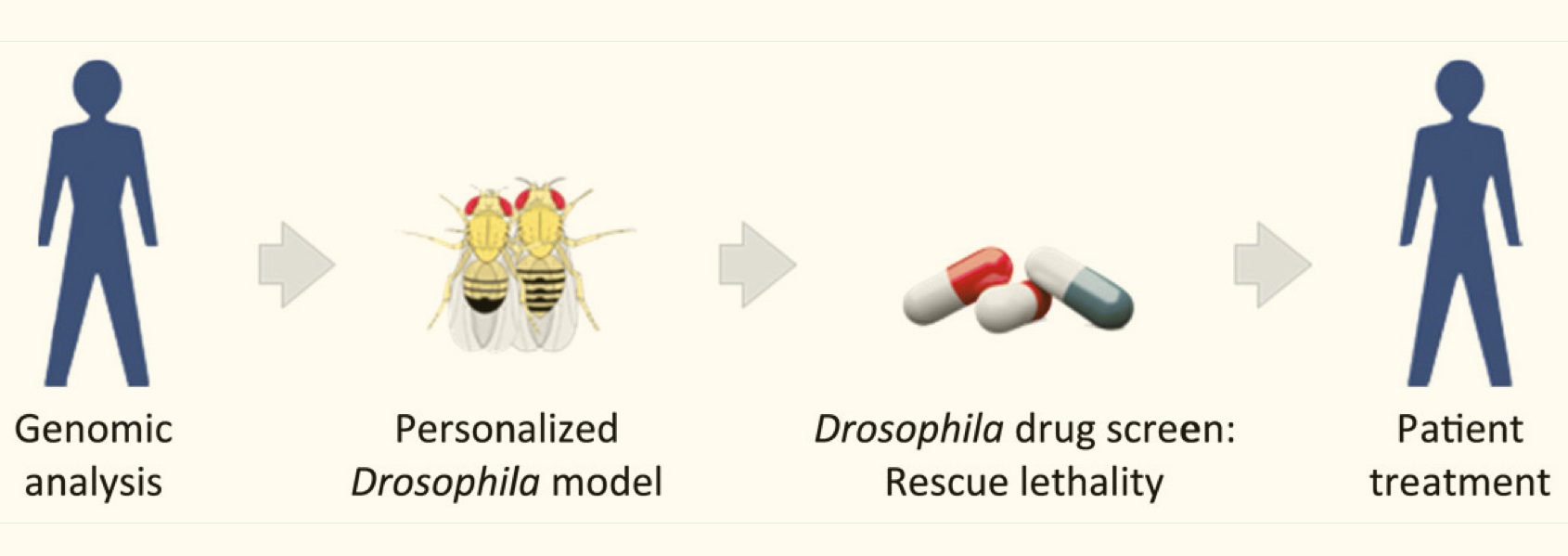
The promise of personalized medicine has been limited by at least two factors: 1) the power of our models—including our ability to get and process data, and 2) our ability to test potential therapeutic solutions in meaningful biological systems, rapidly and systematically, before testing in humans. While we’re seeing rapid and predictable improvements with the power of our models corresponding improvements in how we test therapies have been less consistent. Even if you have a good, genome-level understanding of an individual’s cancer, there is no guidebook for what treatment will be effective against that particular type of cancer. One promising approach has been to use fruit flies (Drosophila) genetically-modified with similar mutational loads as cancer patients to test the efficacy of drug combinations for suppressing tumor growth.

Ross Cagan is an exceptionally creative researcher who leads a lab at the Icahn School of Medicine at Mount Sinai and has been using fruit flies to develop personalized medicine approaches for over 10 years. This week, the Cagan lab published one of the first examples of clinically effective therapies based on such an approach, with lead author Dr. Erdem Bangi (see below for abstract and link). This article describes how Drosophila were screened for effective drug combinations for inhibiting tumors with a similar composition and complexity as a terminally ill colorectal cancer patient’s tumors, and how a specific drug combination was identified and used to effectively shrink the patient’s tumors. Though this treatment did not provide a permanent cure, it did appear to extend the patient’s life and is important proof that this type of approach can be effective.

Excellent, more in-depth summaries can be found HERE and HERE and HERE, so check them out.

The original article, with its somewhat difficult to penetrate title…

Bangi, E., et al. (2019) A personalized platform identifies trametinib plus zoledronate for a patient with KRAS-mutant metastatic colorectal cancerScience Advances

Abstract

Colorectal cancer remains a leading source of cancer mortality worldwide. Initial response is often followed by emergent resistance that is poorly responsive to targeted therapies, reflecting currently undruggable cancer drivers such as KRAS and overall genomic complexity. Here, we report a novel approach to developing a personalized therapy for a patient with treatment-resistant metastatic KRAS-mutant colorectal cancer. An extensive genomic analysis of the tumor’s genomic landscape identified nine key drivers. A transgenic model that altered orthologs of these nine genes in the Drosophila hindgut was developed; a robotics-based screen using this platform identified trametinib plus zoledronate as a candidate treatment combination. Treating the patient led to a significant response: Target and nontarget lesions displayed a strong partial response and remained stable for 11 months. By addressing a disease’s genomic complexity, this personalized approach may provide an alternative treatment option for recalcitrant disease such as KRAS-mutant colorectal cancer.

READ MORE …

Thank you for reading!

Promising result in Cancer Vaccine Clinical Trial

Mount Sinai Researchers Develop Treatment That Turns Tumors Into Cancer Vaccine Factories

Promising result in Cancer Vaccine Clinical Trial

Researchers at Mount Sinai have developed a novel approach to cancer immunotherapy, injecting immune stimulants directly into a tumor to teach the immune system to destroy it and other tumor cells throughout the body. 

The “in situ vaccination” worked so well in patients with advanced-stage lymphoma that it is also undergoing trials in breast and head and neck cancer patients, according to a study published in Nature Medicine in April.

The treatment consists of administering a series of immune stimulants directly into one tumor site.  The first stimulant recruits important immune cells called dendritic cells that act like generals of the immune army. The second stimulant activates the dendritic cells, which then instruct T cells, the immune system’s soldiers, to kill cancer cells and spare non-cancer cells. This immune army learns to recognize features of the tumor cells so it can seek them out and destroy them throughout the body, essentially turning the tumor into a cancer vaccine factory.

“The in situ vaccine approach has broad implications for multiple types of cancer,” said lead author Joshua Brody, MD, Director of the Lymphoma Immunotherapy Program at The Tisch Cancer Institute at the Icahn School of Medicine at Mount Sinai. “This method could also increase the success of other immunotherapies such as checkpoint blockade.”

After testing the lymphoma vaccine in the lab, it was tested in 11 patients in a clinical trial. Some patients had full remission from months to years. In lab tests in mice, the vaccine drastically increased the success of checkpoint blockade immunotherapy, the type of immunotherapy responsible for the complete remission of former President Jimmy Carter’s cancer and the focus of the 2018 Nobel Prize in Medicine.


READ MORE …

Thank you for reading!

Another helpful summary of the recent paper linking jumping genes to cancer

A recent post discussed an important paper that demonstrated an association between transposable elements, aka ‘jumping genes’, and cancer. Transposable elements are an important but often forgotten class of mutagen that can contribute to genome instability and may disrupt genes and their expression. The original article has an outstanding abundance of data and is not easy reading. The article below provides an accessible summary that’s worth reading if you aren’t an expert in genomics.


Cancer: Scientists find 129 'jumping genes' that drive tumor growth

Catharine Paddock, Medical News Today

Another helpful summary of the recent paper linking jumping genes to cancer

In cancer research, scientists usually look for cancer genes by scouring the genome for altered sequences — or mutations — in DNA. But a new study has now revealed that jumping genes, which customary sequencing overlooks, are also important drivers of tumor growth.

Scientists at the Washington University School of Medicine in St. Louis, MO, found that jumping genes are widespread in cancer and promote tumor growth by forcing cancer genes to remain switched on.

They analyzed 7,769 tumor samples from 15 different types of cancer and found 129 jumping genes that can drive tumor growth through their influence on 106 different cancer genes.

The jumping genes were functioning as "stealthy on-switches" in 3,864 of the tumors that the team analyzed. These tumors came from breast, colon, lung, skin, prostate, brain, and other types of cancer.

A recent Nature Genetics paper gives a full account of the study.


Cancer signaling studies take a page from Genetics methods

The following article provides insights into the promise of applying quantitative approaches in the context of tumor tissues and clinical environments. Genetics approaches have dominated cancer research because they generate such an abundance of data ( and because the methodology is so widely and readily generalizable). While genetic variations clearly play an important role in cancer, deviant signaling drives cancer progression and signaling molecules are the targets of most chemotherapeutics. This highlights the importance of understanding cancer signaling pathways with data-rich and quantitatively rigorous methods, similar to those used in genetics. The following article in Science Signaling discusses this topic and is both thorough and accessible. —RPR


Why geneticists stole cancer research even though cancer is primarily a signaling disease

Michael B. Yaffe, Science Signaling

Cancer signaling studies take a page from Genetics methods

Abstract—Genetic approaches to cancer research have dramatically advanced our understanding of the pathophysiology of this disease, leading to similar genetics-based approaches for precision therapy, which have been less successful. Reconfiguring and adapting the types of technologies that underlie genetic research to dissect tumor cell signaling in clinical samples may offer an alternative road forward.


READ MORE …


Thank you for reading!

Chemotherapy and Immune Response, Complex Therapeutic Terrain

Chemotherapy-Stimulated Immune Response: An Open Debate

Jonathan Goodman, Cancer Therapy Advisor

Chemotherapy and Immune Response, Complex Therapeutic Terrain

“A recent review suggested that chemotherapy may prime cancer to respond to checkpoint inhibition.1 According to the review, which was published in the Annals of Oncology earlier this year, this may occur for a variety of reasons, depending primarily on the mechanism of action of the chemotherapy in question. 

In the past, these predictions may have been surprising to researchers in oncology, as chemotherapy was previously thought to be immunosuppressive. Yet, the authors argue, the effects of chemotherapy can “induce favorable immunogenic conditions within the tumor microenvironment, which may be difficult to achieve by just targeting immune cells.” 

In this setting, chemotherapy functions as the first part of a 2-stage evolutionary trap, where in the first stage clinicians actively select for a tumor microenvironment in which checkpoint blockade is most likely to be effective. With cyclophosphamide, for example, immunogenic cell death may be induced, and the drug may lead to dendritic cell homeostasis.2,3 Both are favorable immunomodulatory effects that may lead to an improved immune response —especially, it appears, when checkpoint blockade is used. 

A recent editorial published in the Annals of Oncology, however, suggests that the notion of turning “cold” tumors “hot” may be a misconception.4 This, according to a study author, Thomas Helleday, PhD, professor of translational oncology and director of the Sheffield Cancer Centre at the University of Sheffield, England, is for several key reasons, each of which has to do with the selective processes caused by chemotherapeutics.“


READ MORE …

Gene-Expression Profiling to Understand Cancers of Unknown Origins

Phase 2 Trial Examines Gene-Expression Profiling for Cancer of Unknown Primary Site

A randomized phase 2 trial examining the assignment of treatment based on gene-expression profiling compared with standard chemotherapy for patients with cancer of unknown primary site showed no improvement in the 1-year survival rate with the more tailored approach. However, several caveats may limit the relevance of the findings. A report of this study was published in the Journal of Clinical Oncology.1

Gene-Expression Profiling to Understand Cancers of Unknown Origins

Cancer of unknown primary site (CUP) refers to malignancies in which the originating tumor type cannot be identified. As a result, determining the best treatment for this cancer, diagnosed in approximately 31,000 people in the US each year, is extremely difficult.2 In recent years, oncologists have looked to genetic testing to identify the cancer type as a way to improve care.

In the current study, a molecular analysis of biopsied tissue predicted the originating cancer site for all of the 101 patients treated. The analysis identified a total of 16 sites; cancers of the pancreas (21% of participants), gastric system (21% of participants), and malignant lymphomas (20% of patients) were the 3 most common sites to be predicted as the primary site of malignancy. The Japan-based researchers then randomized the patients to receive therapy appropriate to the predicted site of origin (50 patients) or the standard, empiric treatment of paclitaxel plus carboplatin (51 patients).


READ MORE …

Cancer evading the Immune System, covered in the New York Times

Cancer’s Trick for Dodging the Immune System

Matt Richtel, The New York Times

Cancer evading the Immune System, covered in the New York Times

Cancer immunotherapy drugs, which spur the body’s own immune system to attack tumors, hold great promise but still fail many patients. New research may help explain why some cancers elude the new class of therapies, and offer some clues to a solution.

The study, published on Thursday in the journal Cell, focuses on colorectal and prostate cancer. These are among the cancers that seem largely impervious to a key mechanism of immunotherapy drugs.

The drugs block a signal that tumors send to stymie the immune system. That signal gets sent via a particular molecule that is found on the surface of some tumor cells.

The trouble is that the molecule, called PD-L1, does not appear on the surface of all tumors, and in those cases, the drugs have trouble interfering with the signal sent by the cancer.


READ MORE …

Long non-coding RNA triggers Cancer Resistance

Long non-coding RNA GBCDRlnc1 induces chemoresistance of gallbladder cancer cells by activating autophagy

Qiang Cai, Shouhua Wang, Longyang Jin, Mingzhe Weng, Di Zhou, Jiandong Wang, Zhaohui Tang and Zhiwei Quan

Molecular Cancer (Research Article)

Background

Gallbladder cancer is the most common biliary tract malignancy and not sensitive to chemotherapy. Autophagy is an important factor prolonging the survival of cancer cells under chemotherapeutic stress. We aimed to investigate the role of long non-coding RNAs (lncRNAs) in autophagy and chemoresistance of gallbladder cancer cells.

Methods

We established doxorubicin (Dox)-resistant gallbladder cancer cells and used microarray analysis to compare the expression profiles of lncRNAs in Dox-resistant gallbladder cancer cells and their parental cells. Knockdown or exogenous expression of lncRNA combined with in vitro and in vivo assays were performed to prove the functional significance of lncRNA. The effects of lncRNA on autophagy were assessed by stubRFP-sensGFP-LC3 and western blot. We used RNA pull-down and mass spectrometry analysis to identify the target proteins of lncRNA.

Results

The drug-resistant property of gallbladder cancer cells is related to their enhanced autophagic activity. And we found a lncRNA ENST00000425894 termed gallbladder cancer drug resistance-associated lncRNA1 (GBCDRlnc1) that serves as a critical regulator in gallbladder cancer chemoresistance. Furthermore, we discovered that GBCDRlnc1 is upregulated in gallbladder cancer tissues. Knockdown of GBCDRlnc1, via inhibiting autophagy at initial stage, enhanced the sensitivity of Dox-resistant gallbladder cancer cells to Dox in vitro and in vivo. Mechanically, we identified that GBCDRlnc1 interacts with phosphoglycerate kinase 1 and inhibits its ubiquitination in Dox-resistant gallbladder cancer cells, which leads to the down-regulation of autophagy initiator ATG5-ATG12 conjugate.

Conclusions

Our findings established that the chemoresistant driver GBCDRlnc1 might be a candidate therapeutic target for the treatment of advanced gallbladder cancer.

READ MORE …


Looking Everywhere for Cancer Drugs

Nature’s Bounty: Revitalizing the Discovery of New Cancer Drugs from Natural Products

Cancer Currents Blog

Dr. Grkovic has spent the last several years intimately involved in improving processes for analyzing products of nature—from marine creatures to soil-dwelling fungi to plant leaves—to see whether chemical compounds within them might be starting blocks for new cancer drugs. (The Natural Products Support Group is part of the Frederick National Laboratory for Cancer Research, an NCI-sponsored contractor-operated facility.)

Looking Everywhere for Cancer Drugs

The work has been part of an ambitious, Cancer MoonshotSM-funded initiative, called the NCI Program for Natural Products Discovery (NPNPD), to make it easier for other researchers to mine nature for leads on new cancer drugs.

A big part of that story has taken place in the well-worn home of NCI’s Natural Products Branch, on the NCI campus in Frederick, MD.


READ MORE …

Tasmanian devils adapting to transmissible cancers

Tasmanian devils 'adapting to coexist with cancer'

There's fresh hope for the survival of endangered Tasmanian devils after large numbers were killed off by facial tumours.

The world's largest carnivorous marsupials have been battling Devil Facial Tumour Disease (DFTD) for over 20 years.

But researchers have found the animals' immune system to be modifying to combat the assault.

Tasmanian devils adapting to transmissible cancers

And according to an international team of scientists from Australia, UK, US and France, the future for the devils is now looking brighter.

"In the past, we were managing devil populations to avoid extinction. Now, we are progressively moving to an adaptive management strategy, enhancing those selective adaptations for the evolution of devil/DFTD coexistence," explains Dr Rodrigo Hamede, from the University of Tasmania.

READ MORE …

Clinical application of tumor evolution analysis

Translating insights into tumor evolution to clinical practice: promises and challenges

Matthew W. Fittall and Peter Van Loo

Genome Medicine (Review Article)

Clinical application of tumor evolution analysis

Abstract—Accelerating technological advances have allowed the widespread genomic profiling of tumors. As yet, however, the vast catalogues of mutations that have been identified have made only a modest impact on clinical medicine. Massively parallel sequencing has informed our understanding of the genetic evolution and heterogeneity of cancers, allowing us to place these mutational catalogues into a meaningful context. Here, we review the methods used to measure tumor evolution and heterogeneity, and the potential and challenges for translating the insights gained to achieve clinical impact for cancer therapy, monitoring, early detection, risk stratification, and prevention. We discuss how tumor evolution can guide cancer therapy by targeting clonal and subclonal mutations both individually and in combination…

READ MORE …


Next Generation Sequencing Assay for Blood Cancers

Researchers Develop Targeted Next Generation Sequencing Assay for Myeloid Neoplasms

Researchers from South Korea said they have developed a next-generation sequencing (NGS) assay to detect somatic mutations, translocations, and germline mutations in a single assay for the purpose of supplementing or replacing conventional tests in patients with myeloid neoplasms.

Writing in a recent issue of PLoS One, the researchers said were able to discover a high frequency of germline mutations in cancer predisposition genes. Patients with these mutations exhibited different clinical characteristics, suggesting that germline predisposition has significant clinical implications.

Deep sequencing of Adult Gliomas has promising results

Prospective Clinical Sequencing of Adult Glioma

Siyuan Zheng, Kristin Alfaro-Munoz, Wei Wei, Xiaojing Wang, Fang Wang, Agda Karina Eterovic, Kenna R Mills Shaw, Funda Meric-Bernstam, Gregory N Fuller, Ken Chen, Roel G. Verhaak, Gordon B. Mills, W.K. Alfred Yung, Shiao-Pei Weathers and John F. de Groot

Molecular Cancer Therapeutics (Research Article)

abstract-2105402_640.jpg

Abstract—Malignant gliomas are a group of intracranial cancers associated with disproportionately high mortality and morbidity. Here we report ultradeep targeted sequencing of a prospective cohort of 237 tumors from 234 patients consisting of both glioblastoma (GBM) and lower-grade glioma (LGG) using our customized gene panels. We identified 2485 somatic mutations including single nucleotide substitutions and small indels using a validated in-house protocol. Sixty one percent of the mutations were contributed by 12 hypermutators. The hypermutators were enriched for recurrent tumors, had comparable outcome, and most were associated with temozolomide exposure. TP53 was the most frequently mutated gene in our cohort, followed by IDH1 and EGFR. We detected at least one EGFR mutation in 23% of LGGs, which was significantly higher than 6% seen in TCGA, a pattern that can be partially explained by the different patient composition and sequencing depth. IDH hotspot mutations were found with higher frequencies in LGG (83%) and secondary GBM (77%) than primary GBM (9%). Multivariate analyses controlling for age, histology, and tumor grade confirm the prognostic value of IDH mutation. We predicted 1p/19q status using the panel sequencing data, and received only modest performance by benchmarking the prediction to Fluorescent In Situ Hybridization (FISH) results of 50 tumors. Targeted therapy based on the sequencing data resulted in three responders out of 14 participants. In conclusion, our study suggests ultradeep targeted sequencing can recapitulate previous findings and can be a useful approach in the clinical setting.


READ MORE …

Specificity helps with cancer outcome prediction, therapies

Acute Erythroleukemia Genomic Subtypes Help Predict Outcomes, Suggest Therapies

blood-2194498_640.jpg

NEW YORK (GenomeWeb) – A new genomic analysis of acute erythroid leukemia (AEL) has uncovered recurrent tumor gene mutation and expression profiles, including genomic features that appear to coincide with outcomes for patients affected by the rare, difficult-to-treat form of acute myeloid leukemia (AML).

"These results mark a new era in understanding and treatment of AEL, an aggressive leukemia that has been plagued by diagnostic controversy and poor outcomes," senior author Charles Mullighan, a pathology researcher and co-leader of the St. Jude Children's Research Hospital's hematological malignancies program, said in a statement. 

As they reported online today in Nature Genetics, Mullighan and colleagues performed whole-genome, exome, targeted, and transcriptome sequencing on samples from 159 pediatric or adult AEL patients treated at sites around the world, comparing the somatic mutations and gene expression patterns they found to those in samples from more than 1,900 individuals with non-AEL conditions — from other forms of AML to myelodysplastic syndrome.


READ MORE …

Hope in the fight against Glioblastoma

New Strategies Take On the Worst Cancer—Glioblastoma

““The most common form of malignant brain cancer—called a glioblastoma—is notoriously wily and considered the deadliest human cancer. Glioblastomas charge their way into normal brain tissue diffusely and erratically, making them surgical nightmares. And they mutate at such a rapid rate that most currently available cancer treatments can't keep up with them. Even neighboring tumor cells can be genetically distinct, and therefore hard to target with a single therapy. 

Survival rates from glioblastomas enjoyed a modest bump in the 1980s when radiation became a standard part of the treatment protocol. Patients could expect to live for nearly another year after diagnosis, up from just four to six months. The introduction of the chemotherapy drug temozolomide in the 2000s increased survival another few months. But since then patient survival rates have stalled… many experts insist the key to beating glioblastoma will entail personalizing care to a patient’s individual tumor and the particular molecular signature of a cancer.

One method of testing cancer therapies, including glioblastomas, has been so-called “ex vivo” cancer models, in which malignant cells are probed in the lab. Also known as tumor “avatars,” they allow researchers to test a drug on patients’ cancer cells before introducing it into their bodies. A number of ex vivo models have been tried over the years: culturing tumor cells in Petri dishes; grafting them into animal models; even growing “organoids” (three-dimensional tumors grown on a supporting matrix). These techniques have seen varying success depending on the tumor type at hand, but none have proven especially helpful for glioblastoma.

A report on new research technology published recently in the journal Nature Biomedical Engineering may address the limitations of previous ex vivo approaches. In short, researchers have concocted a glioblastoma-on-a-chip. Chip-based models of various organs and diseases—including many cancers—have debuted in the last few years. They are constructed by lining a plastic microchip with live human cells that mimic a particular organ or disease in order to simplify, cheapen and increase the efficiency of drug testing. The Wyss Institute at Harvard University and other groups have made impressive headway in developing a number of chip-based biologic models. Chip models of the lung, the intestine, skin, bone marrow ALS—even the blood-brain barrier—have been tested.”

READ MORE …

BRCA Challenge creates database for fighting cancer

BRCA Exchange aggregates data on thousands of BRCA variants to inform understanding of cancer risk

A global resource that includes data on thousands of inherited variants in the BRCA1 and BRCA2 genes is available to the public. The BRCA Exchange was created through the BRCA Challenge, a long-term demonstration project initiated by the Global Alliance for Genomics and Health (GA4GH) to enhance sharing of BRCA1 andBRCA2 data. The resource, available through a website and a new smartphone appExit Disclaimer, allows clinicians to review expert classifications of variants in these major cancer predisposition genes as part of their individual assessment of complex questions related to cancer prevention, screening, and intervention for high-risk patients. 

The five-year BRCA Challenge project was funded in part by the National Cancer Institute (NCI), part of the National Institutes of Health, and through the Cancer Moonshot℠. A paper detailing the development of the BRCA Exchange was published January 8, 2019, in PLOS Genetics. READ MORE …

BRCA-challenge-infographic.__v200497217.png


microRNA and cancer therapy

Scientists home in on microRNA processing for novel cancer therapies

microRNA and cancer therapy

“More than a decade of research on the mda-7/IL-24 gene has shown that it helps to suppress a majority of cancer types, and now scientists are focusing on how the gene drives this process by influencing microRNAs. Published this week in the journal Proceedings of the National Academy of Sciences, the findings could potentially have implications beyond cancer for a variety of cardiovascular and neurodegenerative diseases caused by the same microRNA-driven processes.”


READ MORE …